Cryogenic Electron Microscopy uses an electron beam to image samples preserved in a frozen, hydrated state, to reveal the 3D structures of proteins and large molecular complexes at atomic resolution.
Applications:
The Cryo-EM platform proposes different services.
Cryo-EM imaging:
– Cryo-EM grid screening
– Ice thickness
– Particles distribution
– SPA data collection
– 2D classification and 3D cryo-EM map
– Cryo-electron tomography of viruses, bacteria, cell lamella
Negative stain:
– Sample characterisation (proteins/bacteria/viruses)
– Sample quality evaluation for further cryo-EM analysis
– 15-20 A resolution 3D reconstruction of protein/protein complexes
Cellular electron microscopy:
– Observation of sections of embedded samples (bacteria, cells)
– Observation of fixed bacteria, phages, virus
Equipment:
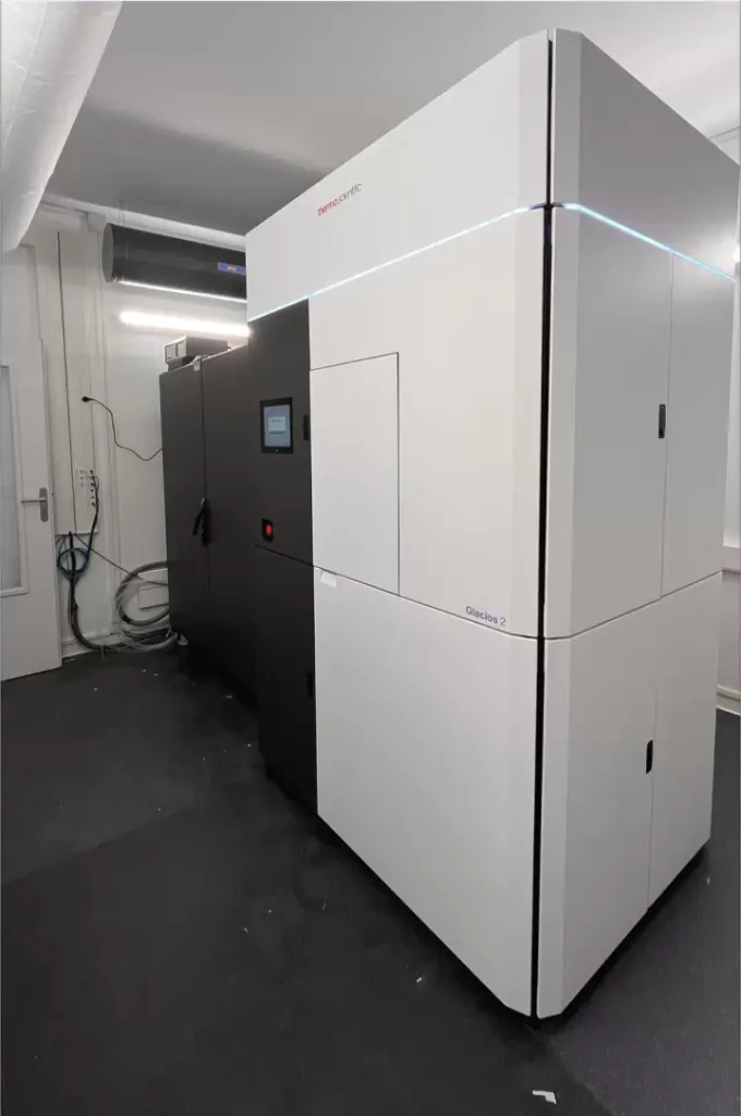
– Glacios 2 200 kV TEM microscope coupled to a Falcon4 Direct electro detector
– Electron microscope JEOL 1400 equipped with a Lab6 operated up to 120 keV.
– EM-GP2 plunger (Leica) to freeze cryo-grids.