The cryo-electron microscopy platform comprises two cryo-electron microscopes 200 kV and 120 kV (Tecnai FEI), each equipped with a direct detection camera (K2 Gatan) and a liquid nitrogen cooled holder (Gatan 626). The 200 kV microscope is equipped with a field emission gun (FEG) and a camera that detects single electrons (Gatan K2 summit); the 120 kV has a LaB6 filament and a basic direct detection camera (Gatan K2 base).
Cryo-electron microscopy visualizes biological objects in their natural aqueous environment. Cold stage stability and the properties of the camera determine the resolution of the structural data obtained (1-0.25 nm).
The platform has a FEG 200 kV cryo-electron microscope and a side-entry stage that is less stable than top entry holders. However, the microscope is equipped with a direct detection camera that, due to its high sensitivity, allows the recording of movies composed of frames with short time exposure (0.1s). Centering and averaging of images from the movie can compensate for the holder drift, leading to high signal to noise ratios. The 200 kV cryo-electron microscope of the platform is thus an ideal tool to initiate studies and to obtain an initial model at a resolution better than 1 nm. Such a model will be very useful to determine the atomic structure of the object with the 300kV microscopes available at Grenoble and Strasbourg. For single objects, the 3D structure can be carried out using tomography at a resolution of a few nm.

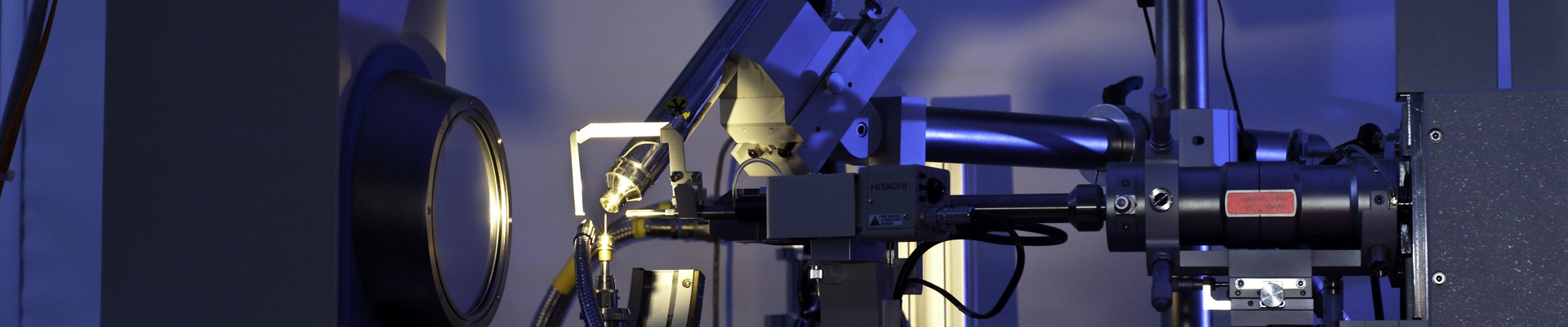
Figure 1: 200kV cryo-electron microscope equipped with a FEG, a liquid-nitrogen
The platform engineers will first test by both conventional and cryo-electron microscopy the feasibility of the potential projects. Those projects which show potential can be continue by the user after they have followed a short introductory course on the 120 kV cryo-electron microscope. In most cases, users should bring samples in a vitrified state. Finally, a few subjects can be taken further in collaboration with the platform engineers.
The platform has only limited computing capabilities, for carrying out preliminary image processing. Where necessary, more extensive processing may be carried out on on external CNRS computers.
The microscopes are installed in a P2 security laboratory, and so projects requiring this security environment can be considered with the agreement of the Scientific committee of the department

Figure 2: 3D reconstruction of a virus at resolution better than 1nm. The atomic model of the capsid protein, determined by X-ray crystallography has been fitted in the map.
Partnership with Genopole
The Genopole has complementary expertise in AFM
see their webpage : http://sabnp.univ-evry.fr/?page_id=43